American Synesthesia
Association
Presentation Schedule and Abstracts
University of California San Diego
Conference Presenters and Abstracts
The 9th Annual National Conference of the American Synesthesia Association took place on October 14 - 16, 2011 at the University of California San Diego. The conference was hosted by V. S. Ramachandran and David Brang of the Brain and Cognition Center.
We were pleased to have Edward M. Hubbard, Post Doctorate Fellow in the Department of Psychology and Human Development at Vanderbilt University, as our Keynote Speaker.
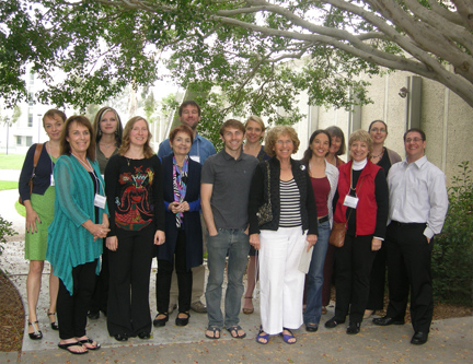
Photo credit: Carter R. Jones
Presenters from left to right: Samantha Moore, Marcia Smilack, Janina Neufeld, Anja Moos, Patricia Lynne Duffy, Marcus Watson, David Brang, Romke Rouw, Greta Berman, Ursina Teuscher, Daphne Maurer, Carol Steen, Lilach Akiva-Kabiri, Edward M. Hubbard. Not present in photo: Patricia Albers, Helena Melero, V.S. Ramachandran, Mark Stewart, Steffie Tomson.
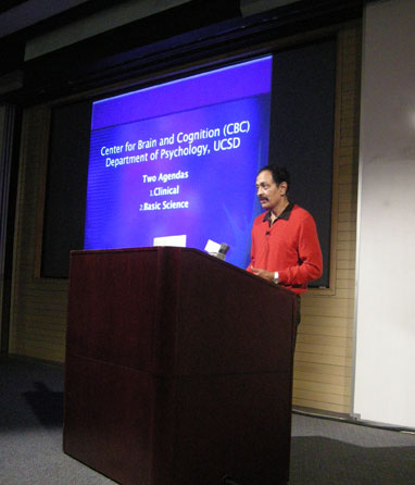
Photo credit: Carter R. Jones
V.S. Ramachandran
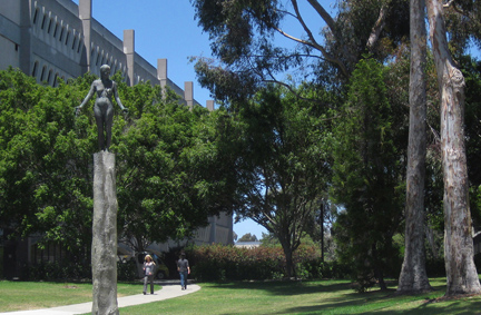
UCSD Campus, Photo Credit: Michael Ghiam
Presentation Schedule
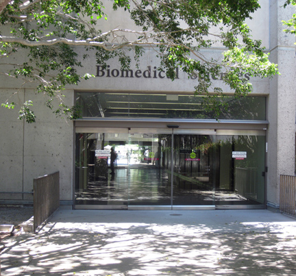
Held in Garren Auditoruim in the Biomedical Sciences Building
October 14 – 16, 2011
Abstracts
Lilach Akiva-Kabiri, Ben-Gurion University of the Negev, Israel
Music Pitch Tone Synesthesia: general and specific aspects
Lilach Akiva-Kabiri, Limor Gertner & Avishai Henik, Department of Psychology, and the Zlotowski Center for Neuroscience, Ben-Gurion University of the Negev, Beer-Sheva, Israel
Pitch-tone synesthesias are often associated with Absolute Pitch (AP), which is the ability to identify pitch tones without an external tonal context. We tested the importance of absolute pitch (AP) in tone–color (TCS) and tone-space synesthesias (TSS). In TCS, pitch chroma (e.g., Sol) elicits a color perception. In a TSS, musical tones are organized explicitly in a well-defined spatial array. AP and non-AP TCS were presented with a Stroop like task and were asked to name a colored patch on the screen and ignore a musical tone. The synesthetic color elicited by the tone could be congruent or incongruent with the colored patch. When musical tone was auditory, only AP possessors presented a congruency effect, whereas when tone was presented visually, both groups presented a significant congruency effect. In the second part of this work, we used a cue detection task were TSS without AP and controls were asked to detect a visual cue, ignoring a simultaneous irrelevant auditory tone. Synesthetes were slower when auditory tone and cue were incongruent with each other compared with the congruent condition. Hence they were unable to suppress the orientation of the attention to the auditory tone space form even though they did not possess AP. The results of these experiments demonstrate the automaticity and authenticity of these types of synesthesia. Moreover, data suggest that AP modulated effects of TCS but not of TSS. Results are interpreted considering the underlying characteristics of color perception - that is essentially categorical in nature - compared with the more ordinal nature of space.
Patricia Albers, Writer, Curator; Mountain View, California
Joan Mitchell, Artist and Synesthete: Three Paintings
Artist Joan Mitchell (1925-1992) had four types of synesthesia: colored graphemes, colored sound, colored personalities, and emotionally mediated synesthesia, plus eidetic memory. Although she was apparently unaware that synesthesia is a named, normal, and shared condition, Mitchell was acutely conscious of her perceptual gifts and intentional about using them in her art-making. I propose to present and analyze three Mitchell oil paintings, Hemlock (1956), My Landscape II (1967), and Faded Air I (1985), giving primary consideration to the role the artist’s synesthesia may have played in her aesthetic decision-making. Using both verbal and visual evidence, I will examine her choice of subject, medium, and format and use of formal elements (color, shape, line, texture, and light and dark) as they relate to her synesthesia and eideticism and to the Kluver Form Constants.
In addition, I will address Mitchell's practice of abstraction. Considered an Abstract Expressionist and self-identified as an abstractionist, the artist drew upon the theories of psychologist Anton Ehrenzweig regarding the dynamics of a kind of syncretic vision that “ outstrips the powers of conscious attention.” At the same time she explained that she worked from a piece of life “a real image” in which ldquo;motion is made still, like a fish trapped in ice. It is trapped in the painting. I do not conceive.” In what sense, then, are Hemlock, My Landscape II, and Faded Air I abstract works?
Greta Berman, The Juilliard School, New York City
Vincent Van Gogh, Synesthete
Today, more than 100 years after Van Gogh’s tragic and untimely death in 1890, his art and life continue to inspire research. Art historians and psychologists have spent decades attempting, only minimally successfully to understand him. Fortunately, he himself wrote thousands of letters. Most of these have been archived, providing direct evidence of his insights and problems.
Though he may have suffered from one or another mental/emotional malady, several years of studying shared characteristics present in the work of known and acknowledged synesthetes have convinced me that Van Gogh was synesthetic. Indeed, the lack of comprehending this by others and even himself, may have contributed to pushing him over the edge.
In his writing, he often referred to colors having emotions; he described nature in anthropomorphic terms (what we now call personification). Occasionally he mentioned that colors had musical sound for him, and there is the famous story of how his piano teacher threw him out for insisting that the notes he played had colors.
Visual evidence is plentiful, including his use of paint direct from the tube - in a kind of frenzy; his repeated zigzags, wavy lines, and what we now recognize as Kluver’s form constants, as well as his infinite variation of hues.
This paper will explore some of the manifestations of his synesthesia, both in his painting and in his writing. Our awareness today of many more kinds of synesthesia than ever before helps in discerning this.
David Brang, Department of Psychology, University of California, San Diego
Non-synesthetes trained on grapheme-color associations show unconscious priming of color words
David Brang, V.S. Ramachandran, Seana Coulson, Department of Psychology, University of California, San Diego
Grapheme-color synesthesia is defined by subjects’ automatic and consistent experience of colors when viewing achromatic numbers and/or letters. While these perceptions are vivid and perceptually salient, a long debate has raged asking whether synesthesia requires consciousness. In 2001, Mattingley et al. described a behavioral priming paradigm testing whether unconsciously processed graphemes (via a backward masking paradigm) evoke synesthetic colors. Critically, the authors found no effect of priming in synesthetes suggesting that synesthesia requires consciousness. However, this paradigm may have lacked the sensitivity required to detect small effects of unconscious synesthetic processing as behavioral priming must be robust. Using event-related potentials (ERPs), a measure of electrical activity in the brain, researchers have demonstrated unconscious processing of numbers and words in the general population as this technique has the excellent sensitivity for subtle activation in the brain. As an initial control study to test for the presence of unconscious priming in synesthesia, 17 non-synesthetes were trained on 6 grapheme-color associations, and were subsequently testing using an ERP masked priming paradigm. Subjects were presented with either masked (not consciously recognizable) numbers or color words, followed by visible color words. Intriguingly, non-synesthetes showed significant priming effects (indexed by the N400 ERP component) for both masked numbers that were merely associated with particular colors and masked color words. These results show that controls simply trained on number-color pairings show unconscious processing of synesthetic associations, pressing the question of whether synesthesia is processed unconsciously, and if so, is it meaningful in light of the present findings?
Patricia Lynne Duffy, Author; United Nations Language and Communications Programme, New York City
Why do synesthesia authors like “blue”?
Quite a few of the last decade’s books on synesthesia (whether scientific, personal, historical or fictional) have had the word, “blue&drdquo; in their titles: in order of publication, Blue Cats and Chartreuse Kittens, by Patricia Lynne Duffy (2001), The Sound of Blue, by Holly Payne (2005), Born on a Blue Day by Daniel Tammet (2006), The Frog Who Croaked Blue by Jamie Ward (2008); Wednesdays are Indigo Blue by Richard Cytowic and David Eagleman (2009). Through citing contemplations on the color blue by author William Gass and others, the presenter will explore meanings of and associations with the color, “blue”, along with possible reasons why synesthesia authors have chosen to include the word, “blue” in their book titles.
Edward M. Hubbard, Department of Psychology and Human Development, Vanderbilt University, Nashville, Tennessee Keynote Speaker
The Cross-Activation Theory at Ten: Substantial Growth, Future Challenges
In 2001, Ramachandran and Hubbard introduced the cross-activation model of grapheme-color synaesthesia. On the occasion of its tenth birthday, I will review the evidence from experiments that have been conducted to test the model, in order to assess the growth and abilities of the model. I will review data from behavioural, functional neuroimaging (fMRI), anatomical (DTI and VBM) and electroencephalography (EEG) and magnetoencephalography (MEG) studies of grapheme-color synesthesia. Although much of this evidence has supported the basic cross-activation hypothesis, our growing knowledge of the neural basis of synaesthesia, grapheme-processing and colour-processing has necessitated two specific updates and modifications to the basic model: (1) Our original model assumed that binding and parietal cortex functions were normal in synesthesia; we now recognize that parietal cortex plays a key role in synesthetic binding, as part of a two-stage model. (2) Based on MEG data we have recently collected demonstrating that synesthetic responses begin within 140 ms of stimulus presentation and an updated understanding of the neural mechanisms of reading as hierarchical feature extraction, I will present a revised and updated version of the cross-activation model, the cascaded cross-tuning model. I will then briefly discuss data demonstrating that the cross-activation model may be extended to account for other forms of synesthesia, and end with a discussion of open questions about how learning, development and cortical plasticity interact with genetic factors to lead to the full range of synesthetic experiences. An explicitly neurodevelopmental approach will be required to better understand how experience shapes the brain circuits that mediate synesthetic experiences, and may constitute the major challenge for the next ten years of the cross-activation theory.
Daphne Maurer, McMaster University, Ontario, Canada
Sound Symbolism: The Mapping of Sound to Shape in English-learning Children
Daphne Maurer, Ferrinne Spector, Catherine T. Best, University of Western Sydney, Australia
Learning language involves the mastery of arbitrary connections between objects and combinations of sounds. However, some common mappings appear not to be completely arbitrary but to make use of cross-modal connections that underlie some forms of synaesthesia and that are readily understood by non-synaesthetic adults (e.g., Ramachandran & Hubbard, 2001). Here we investigated the influence of such sound symbolism on children, aged 30-36 months, who are in the midst of rapidly acquiring new vocabulary. In Experiment 1, there were four pairs of nonsense words in which one word contained the rounded vowel /o/ and the other word contained the nonrounded vowel /i/ (as in feet) (e.g., /koko/ versus /kiki/). In the context of a game, toddlers matched the words to contrasting pairs of rounded and jagged nonsense shapes. Toddlers (n= 40) consistently matched rounded vowels to rounded shapes and non-rounded vowels to jagged shapes (p < .001). In Experiment 2, there were four contrasting pairs of nonsense words differing in stop versus approximant consonants (e.g., /bibi/ versus /lili/, respectively). Toddlers (n=40) matched consonant sounds to shape randomly, as did 10 adults. In ongoing work, we are investigating other consonant contrasts and the influence of the vowel’s sound (acoustics) versus the shape of the lips when pronouncing it. The results confirm that there are naturally biased correspondences between vowel sound and shape that may influence the child’s learning of vocabulary but have thus far failed to reveal similar influences for consonants.
Helena Melero, Department of Psychobiology, Faculty of Psychology, Universidad Complutense de Madrid, Spain
Achromatic Synesthesia: a combined neurophenomenological and neuroimaging approach
Melero, H.1,2, Peña-Melián, A.3, Ríos-Lago, M.4,5,6 and Álvarez-Linera, J.6,7
1Department of Psychobiology, Faculty of Psychology, Universidad Complutense de Madrid, Spain
2 Department of Investigation, Development and Promotion, International Artecittá Foundation, Spain
3 Department of Anatomy and Embryology of Human Nervous System I, Faculty of Medicine, Universidad Complutense de Madrid, Spain
4 Department of Basic Psychology II, UNED, Madrid, Spain
5 Brain Damage Unit, Beata Mariana Hospital, Madrid, Spain
6Fundación Cien-Fundación Reina Sofía, Madrid, Spain
7 Neuroradiology Section, Department of Radiology, Hospital Ruber International, Madrid, Spain
Grapheme-color is one of the most studied variants of synaesthesia. Most synesthetes report having one or more achromatic grapheme among their letters and numbers, however, to our knowledge, researchers have not focused attention on these black, white and grey experiences.
For the first time, we have carried out an MRI study on achromatic synesthesias for graphemes. Eight associator grapheme-colour synesthetes and matched controls underwent fMRI scans on a 3.0 T Signa HDx MR scanner (GE Healthcare). Neuroimaging data were analysed using SPM5 (Welcome Department of Imaging Neuroscience, London). Qualitative information about synesthetic experiences was taken into account to fully understand the observed brain activity.
The achromatic synesthetic brain web includes bilateral insula, left anterior cingulate cortex, right superior temporal gyrus and left caudate tail. Our results suggest that emotion plays a key role, and could explain not only colour appearance, but also the conscious experience of synaesthesia from an integral multi-dimensional perspective.
Samantha Moore, Director; Shropshire, England
Animation: the synaesthetic art?
Is animation a significant medium for conveying synaesthesia? Animator Samantha Moore will look at how she and other artists have used the medium of animation to convey the intricacies of audio-visual synaesthesia. Is animation a useful tool in communicating synaesthesia and why would this be? What are some of the opportunities and restrictions of using animation to convey synaesthesia? Animation can naturally make apparently surreal links between objects and places them in time and space for the audience using metamorphosis, movement and sound. It can replicate the impression of photo-realism, but it can also convey what Chris Landreth (director of the 2004 academy award winning animated short Ryan) called “psycho-realism” too, “to expose the realism of the incredibly complex, messy, chaotic, sometimes mundane, and always conflicted quality we call human nature.” By using animation synaesthesia can be literally interpreted and visualized for a non-synaesthetic audience, to whom without context it may seem to be entirely abstract. From Allegretto and Fantasia to her own and other contemporary work she will try and unpick why synaesthesia may have a particular affinity for animation, and vice versa.
Anja Moos, University of Glasgow, Scotland
Perception of voice quality by synaesthetes, phoneticians and controls
Anja Moos1, 2, Rachel Smith1, David Simmons2
1Laboratory of Phonetics, School of Critical Studies, University of Glasgow
2School of Psychology, University of Glasgow
Although synaesthetic perceptions triggered by the sounds of people’s voices have been described anecdotally, this sub-type of synaesthesia has received little scientific attention. We therefore conducted an online survey of voice ‡ colour/texture synaesthesia. Voice synaesthetes, phoneticians and controls were asked to describe recorded voices in their own words, choose a colour and a texture which they thought fitted the voice best, and choose the strength of various descriptive attributes (e.g. rough vs. smooth, grey vs. colourful). Two sentences spoken by two trained phoneticians in 10 different voice qualities (VQs) each, e.g. nasal, whisper, falsetto, served as stimuli. There was a short voice identification task at the end.
It was found that pitch influenced brightness and colour associations with the voice for all groups but showed some idiosyncratic patterns for synaesthetes and others for phoneticians. Synaesthetes were less influenced by pitch in their colour associations than others and phoneticians were more systematic than others in their use of scales for high-low and tense-relaxed according to the perceived pitch and VQ. Texture and voice associations will also be presented, as will results from a re-test for consistency. Analysis of the verbal descriptions showed a strong usage of personality descriptions across all groups. Additionally, phoneticians used more technical terms to describe the VQs, but synaesthetes used more terms for colour, texture and shape. Overall, synaesthetes performed less well in the voice identification task than others, but were outstanding in identifying the correct voice in whisper.
Janina Neufeld, Centre of Systems Neuroscience, Department of Psychiatry, Hannover Medical School,
Multimodal integration in grapheme-colour and auditory-visual synaesthetes investigated with the double-flash illusion
Neufeld J, Sinke C, Emrich HM, Zedler M, Bleich S, Szycik GR, Hannover Medical School, Clinic for Psychiatry, Social Psychiatry and Psychotherapy
There is evidence that auditory-visual synaesthesia shares at least some mechanisms with audio-visual integration (Ward et al. 2006). But is enhanced multimodal integration restricted to inducer-concurrent couplings in synaesthesia or are there general differences in this respect between synaesthetes and non-synaesthetes? To address this question grapheme-colour and auditory-visual synaesthetes were investigated by using the double flash illusion paradigm, in which single light flashes together with multiple beep sounds are illusionary perceived as multiple flashes (Shams et al. 2000). By varying the separation of auditory and visual stimuli systematically, the hypothesis of widened temporal window of audio-visual integration in synaesthetes was tested, as resulting in a larger percentage of illusionary perceived flashes in conditions with larger audio-visual separation (more than 100 ms). The results indicate that there are differences between synaesthetes and controls – not in the width of the temporal window but in the number of perceived illusions in trials with short audio-visual separation up to 150 ms.
V.S. Ramachandran Director, Center for Brain and Cognition, University of California San Diego
Synesthesia; sensory cross activation, intersensory abstraction and the big bang of metaphor and cognition
Romke Rouw, University of Amsterdam, Department of Psychology, Brain and Cognition, The Netherlands
Connectivity in synesthesia
One explanation of the ‘crossing-over’ of one sensation into another in synesthesia is that there is literal ‘crossing-over’ of activation from one brain area into another. In line with this model, evidence for increased connectivity has been obtained, as synesthetes show differences in their white matter properties. In this presentation, I will present recent findings on structural as well as functional connectivity studies revealing differences between synesthetes and non-synesthetes. One way to study functional connectivity is by measuring EEG, and map out the electrophysiological dynamics evoked by achromatic grapheme perception in synesthesia. We found that, in particular, projector synesthetes differ from non-synesthetes in alpha and theta power dynamics as well as in inter-site phase synchrony. Results in this study indicate different oscillatory dynamics in the associator versus projector synesthetes. MRI studies show how a network of brain areas underlies synesthesia. The networks involve both sensory-specific brain regions and ‘higher’ or associative brain areas. I will briefly discuss the possible role of these different brain areas in the networks involved in synesthesia.
Marcia Smilack, Reflectionist, Photographer, Writer; Columbus, Ohio
The Perceptual Purdah
I think of synesthesia as a perceptual purdah, a permeable curtain or sheath that is essential to the multi-layering of consciousness but which in non-synesthetes is hidden behind what is presumably a bearing wall. Conversely, awareness of one's synesthesia amounts to removing the first layer often encapsulated in an early memory (for me, it was the first time I struck a piano key and saw green). I sometimes wonder if the difference between synesthetes and non-synesthetes is merely a thin veil of unawareness.
I photograph reflections on the surface of water. I rely on my synesthetic responses to inform me of the decisive moment. I watch the moving water until I hear a chord of color, feel texture against my skin or experience the taste ice cream (to name only three responses); in each case, at that precise moment, I click the shutter. This is how I taught myself photography.
I have other forms of synesthesia. For instance, I see time in topographical maps and I have a tendency to personify virtually everything I see including numbers and letters. I live my life in metaphor, accompanied by what I think of as my doppelganger, the one inside - the navigator - who spots synesthetic events faster than I could or notice them if I tried. Traveling with me on its parallel paths, my synesthetic responses are no doubt responsible for my eidetic memory, the result of automatic double or triple coding. The goal of my lecture in San Diego is to pull aside the perceptual purdah to show you what lies on the other side.
Mary Jane Spiller, School of Psychology, University of East London, UK
Exploring synaesthetes’ mental imagery abilities across multiple sensory modalities with subjective and objective measures
Mary Jane Spillera, Clare Jonasa, Zeeshan Syeda, Julia Simnerb and Ashok Jansaria
aSchool of Psychology, University of East London, UK
bDepartment of Psychology, University of Edinburgh, UK
Previous research on the mental imagery abilities of synaesthetes has concentrated on visual and spatial imagery in synaesthetes with spatial forms (Price, 2009; 2010; Simner et al, 2008) and letter-colour synaesthesia (Spiller & Jansari, 2008). Though Barnett and Newell (2008) asked synaesthetes of all types to fill out a questionnaire on visual imagery, most of their synaesthetes reported some form of linguistic-colour synaesthesia. We extend the investigation of mental imagery to a wider variety of synaesthesia types and a wider variety of sensory modalities using a questionnaire study and several tests of visual and auditory mental imagery ability. Our results indicate that, as a group, synaesthetes self-report greater vividness of visual, auditory, tactile, and taste imagery (but not olfactory, somatic or kinaesthetic imagery) than do non-synaesthetes. They also report making greater use of mental imagery than non-synaesthetes, in everyday activities. In contrast, there is not such a contrast in the synaesthetes’ and non-synaesthetes’ performance on the imagery tests. These results have important implications for our understanding of synaesthesia, and potential fundamental differences in perceptual processing of synaesthetes and non-synaesthetes.
Carol Steen, Artist; Touro College, New York City
Evidence of Synesthesia in 2D - Painting, Photography, and Video, and
3D - Sculpture and Architecture
Synesthesia has a way of appearing in the artworks of those who have it, and commonalities can be found if one knows what to look for. Thanks to the work of Heinrich Kluver and his taxonomy of perceptions that he called Form Constants, researchers in the arts have started to study artworks in terms of aesthetics and processes of art that have previously been unknown to them. They are finding commonalities and interest in the art world is considerable as evidenced by several recent museum exhibitions that have shown the artworks of artists whom curators now believe to have been synesthetic. Artists and art historians have compared the types of shapes used, the spatial arrangements and organization, the depictions of movements and are exploring the reasons for the aesthetics in these artworks. However, until now, almost all the studies have been of two-dimensional artworks, paintings, photographs, and video. I believe the same Form Constants can be used to investigate and discover synesthesia in the works of three-dimensional art. In my paper I will compare my findings of synesthesia in two-dimensional art and explore the question of how synesthesia is used to create three-dimensional works, both sculpture and architecture.
Mark Stewart, Department of Psychology, Willamette University, Oregon
When January is Better Seen Upside Down and to the Right: A Vantage Point Preference for a Time-space Synaesthete
Michelle Jarick, Department of Psychology, University of British Columbia, Canada; Daniel Smilek, Department of Psychology, University of Waterloo, Ontario, Canada; Michael Dixon, Department of Psychology, University of Waterloo, Ontario, Canada
Time-space synaesthetes ‘see’ time units organized in a rigid, idiosyncratic spatial form.
While the structure might be invariant for most synaesthetes, the perspective by which
some view their spatial form can be more flexible. Depending on the synaesthetes’ needs,
some are able to zoom-in and out of their month-space, or change month positions as the
year progresses. One synaesthete (L) adopts different viewpoints dependent on whether a
month is seen or heard. Given a spatial-cueing task with central month names as cues, L
detected targets on the left faster following the written month January, yet faster on the
right following the spoken month January. Curiously, L indicates a preference for her
auditory perspective, even though the month names are represented upside down. To
verify this, we conducted a spatial-cueing task that included visual, auditory, and
audiovisual month cues. The audiovisual cues were congruent with L’s viewpoint
(auditory + month inverted) or incongruent (auditory + month upright). Our key
prediction was that L would show enhanced cueing-effects for the audiovisual congruent
cues - both triggering the auditory perspective – and little or no cueing-effects for the
audiovisual incongruent cues - each triggering opposite viewpoints. As predicted, L
detected targets following the audiovisual congruent cues the fastest (and consistent with
her auditory perspective), while the audiovisual incongruent cues showed smaller cueing effects
(and were consistent with her visual perspective). Here, we demonstrate that not
only can L change perspectives within her time-space, she has a clear preference for the
auditory one.
Ursina Teuscher, Psychology Department, Portland State University, Oregon
Brain responses (fMRI) to month words in time-space synesthetes and controls
David Ruhl, Arvind Caprihan, and Bob Thoma, Mind Research Network (MRN), Albuquerque, New Mexico
This study investigates the synesthetic experience some people have of temporal sequences (e.g., months of the year) being linked to specific spatial locations. The goal of our research is to find out which brain areas are involved in the cross-activation between temporal and spatial processing in synesthetes. With fMRI and DTI measures, we are testing the hypothesis that in this type of synesthesia there may be unusual connectivity in the parietal cortex. This talk will focus on fMRI findings.
10 time-space synesthetes and 15 controls participated in an fMRI experiment. We used a block design, where blocks of month words that presumably elicit a spatial association in synesthetes alternated with blocks of control words. The control words included four separate blocks of spatial and non-spatial nouns and adjectives. The words in each control block were matched to the month words in number of syllables and spoken word frequency.
We found that synesthetes showed overall greater brain activation when listening to month words than did controls. This was the case both when the activation was compared to blocks of resting state and to blocks of control words. Of the brain areas that were activated more strongly in synesthetes than controls, the largest and most significant ones were in the precuneus, an area which is known to be involved in spatial attention and manipulation of mental images, and in other areas of the parietal and frontal lobes.
Steffie Tomson, Baylor College of Medicine, Department of Neuroscience, Houston, Texas
Functional connectivity MRI and family linkage analysis of synesthesia.
S. N. TOMSON1, M. BRAY2, R.A. GIBBS3, S. LEAL3, D. M. EAGLEMAN1;
1Department of Neuroscience, 2 Department of Pediatrics, 3 Department of Molecular and Human Genetics, Baylor College of Medicine
One common form of synesthesia is characterized by an automatic perception of color in response to members of over-learned sequences such as letters, numbers, weekdays, or months. We call this form colored sequence synesthesia (CSS). To elucidate the neural activity underlying CSS, we used neuroimaging to localize grapheme-sensitive brain regions in 17 synesthetes and 17 controls. A contrast of graphemes with scrambled graphemes revealed regions in right and left temporal lobes that are similar in the synesthetic and control groups, with the difference that synesthetic grapheme activity bleeds into the right fusiform gyrus, overlapping the traditional color region, V4. In the second step of the neuroimaging, participants listened to a dynamic presentation of graphemes in the form of audio clips from children’s television. By performing functional connectivity analyses with several seed regions, we found that the grapheme regions in synesthetes were most negatively correlated with color regions in the fusiform gyrus, unmasking a novel relationship between the grapheme and color regions in synesthetes. We are currently using dynamic causal modeling to probe the directional relationship between these seed regions, while using graph theory to examine whole-brain networks that are involved in processing the audio clips. Finally, we are searching for the genetic basis of CSS. We present data from our ongoing family linkage analysis (Tomson et al. 2011), which implicate a region on chromosome 16 containing over 100 genes expressed in the brain. We are in the process of sequencing all family members for a subset of these genes. In summary, we are combining neuroimaging studies and genetic linkage analyses to build a richer picture of the neural basis of synesthesia, an understanding which will serve as a guide to the normal and abnormal operations of neural cross-talk.
Marcus Watson, Department of Psychology, University of British Columbia, Canada
Preliminary Report from the Prague Synaesthesia Survey
Marcus R Watson - Department of Psychology, University of British Columbia, Vancouver, BC, Canada
Jan Chromy - Institute of Czech Language and Theory of Communication, Charles University, Prague, Czech Republic
Lyle Crawford - Department of Philosophy, Simon Fraser University, Burnaby, BC, Canada
David Eagleman - Department of Psychiatry, Baylor College of Medicine, Birmingham, AL, USA
Kathleen Akins - Department of Philosophy, Simon Fraser University, Burnaby, BC, Canada
We present results from the largest survey to date on synaesthesia. As part of a large-scale, cross-linguistic study, we asked 5,001 students at Charles University in Prague about their synaesthetic tendencies. Self-reports of colour synaesthesias (e.g. letter-colour, weekday-colour, etc) were tested using the online Synesthesia Battery website (Eagleman et al., 2007). Results show that there are at minimum 3.2% colour synaesthetes in the general population. We also show that gender differences in reported rates of synaesthesia can be explained by response bias, as hypothesized by Simner et al. (2006). Women were more likely than men to respond positively to the survey questions, and more willing to comply with subsequent requests for further participation, but were no more likely to be confirmed synaesthetic by the consistency tests than their male counterparts. We also discuss “less-than-perfect” synaesthesias, in which participants show highly consistent colour associations for only some inducers, e.g. grapheme-colour synaesthetes with only 12 consistently–coloured letters. Finally, we present the curious phenomenon of individuals who have extremely consistent colours for letters, weekdays, or months, but were apparently unaware of this fact prior to the survey. Between 10 and 20% of all the synaesthetes in our sample showed this peculiar trait. The implications of all these results for synaesthesia theory and research will be discussed.